
Can an energy-based modality safely replace cold steel for delicate mole removal without trading a surgical scar for erratic thermal damage? For clinicians, selecting a professional plasma pen for benign lesions involves critical concerns regarding post-inflammatory hyperpigmentation, healing predictability, and Health Canada compliance. The proliferation of low-voltage, unregulated spark devices has blurred the line between cosmetic novelties and certified medical-grade systems.
You already recognize that tissue preservation isn’t optional when treating pigmented lesions, particularly on visible areas where patient expectations centre on flawless cosmesis. Discover how professional-grade direct current plasma technology provides the precision required for safe, effective benign mole removal in a clinical setting. This guide examines the biophysics of direct current sublimation, reviews pre-procedural dermoscopic screening rules, and outlines the regulatory pathways for Canadian medical practices.
Key Takeaways
- Deploying a professional plasma pen for benign lesions leverages controlled atmospheric sublimation rather than mechanical excision, vaporizing targeted tissue without surgical sutures.
- Patented Direct Current (DC) technology delivers a continuous 0.1 mm focal discharge that minimizes lateral thermal spread compared to erratic alternating current spark devices.
- Comprehensive dermoscopic screening is mandatory prior to ablation, as the complete sublimation of tissue precludes subsequent histological evaluation.
- Canadian clinicians must operate Health Canada licensed Class III medical devices backed by MDSAP certification to maintain regulatory compliance and clinical safety standards.
- Stable direct current energy accelerates re-epithelialization, promoting predictable carbon crust shedding within three to seven days for optimized aesthetic outcomes.
The Clinical Role of Plasma Technology in Benign Mole Removal
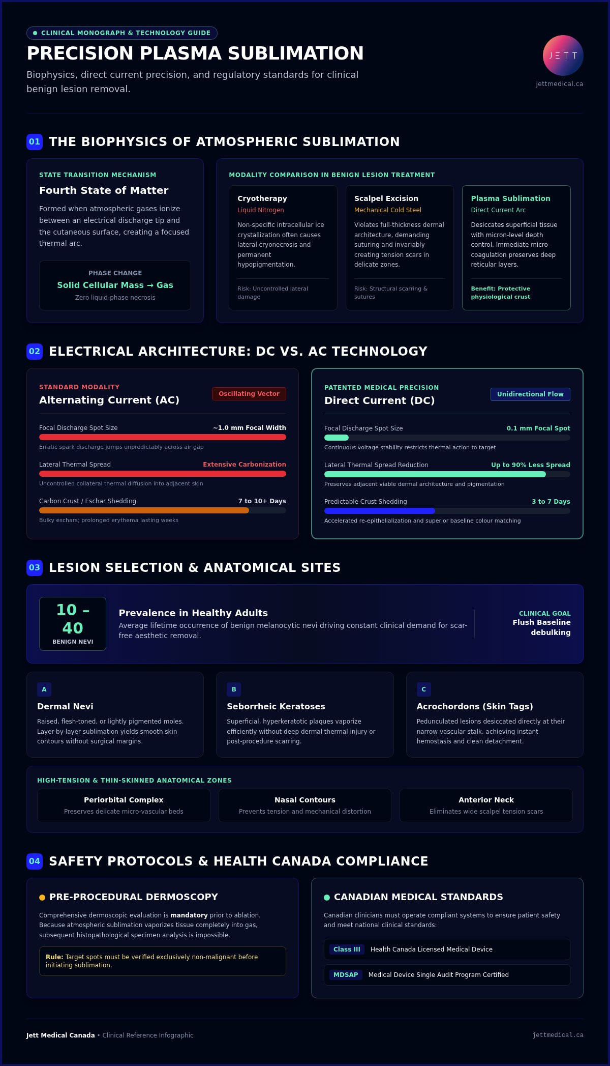
Medical plasma represents the fourth state of matter, formed when atmospheric gases ionize between an electrical discharge tip and the cutaneous surface. This electrostatic phenomenon creates a focused thermal arc that vaporizes targeted epidermal structures through sublimation, transitioning solid cellular mass directly into gas without liquid-phase necrosis. Utilizing a professional plasma pen for benign lesions allows practitioners to desiccate superficial tissue with micron-level depth control, triggering immediate micro-coagulation without disturbing deeper reticular layers.
How Plasma Sublimation Differs from Traditional Methods
Traditional destruction modalities carry distinct clinical disadvantages. Cryotherapy utilizes liquid nitrogen to induce non-specific intracellular ice crystallization, often causing lateral cryonecrosis and permanent hypopigmentation. Surgical scalpel excision, while diagnostic, violates full-thickness dermal architecture, demanding suturing and invariably creating tension scars. Plasma sublimation functions as an energetic shave, debulking elevated tissue layer by layer down to the flush baseline. Surrounding healthy skin remains thermally protected, which prevents wide scar formation and facilitates primary intention re-epithelialization beneath a protective physiological crust.
Identifying Suitable Benign Lesions for Treatment
Clinical efficacy hinges on precise lesion selection. Ablative technologies eliminate the cellular specimen, removing any opportunity for subsequent histopathological analysis. Consequently, clinicians must verify that targeted spots are exclusively non-malignant.
- Dermal Nevi: Raised, flesh-toned, or lightly pigmented moles present ideal substrates for layer-by-layer sublimation, yielding smooth skin contours without scalpel margins.
- Seborrheic Keratoses: Superficial, hyperkeratotic plaques vaporize efficiently without deep dermal injury.
- Acrochordons (Skin Tags): Pedunculated lesions can be desiccated directly at their narrow vascular stalk with instant hemostasis.
Because healthy adults display between 10 to 40 benign melanocytic nevi across their lifetime, patient demand for cosmetically superior removal remains constant. A medical-grade plasma system proves particularly advantageous across anatomically delicate regions such as the periorbital complex, nose, and anterior neck. In these high-tension or thin-skinned zones, cold steel excisions often yield unacceptable cosmetic tension, while plasma delivers pinpoint energy that preserves delicate micro-vascular beds.
DC vs. AC Plasma: Why Precision Matters for Pigmented Lesions
In clinical dermatology, the electrical architecture powering the device dictates procedural safety and tissue response. Alternating current (AC) systems generate oscillating electrical arcs that jump unpredictably across the air gap between the probe and the skin. This oscillation creates an erratic spark discharge with a focal width of roughly 1.0 mm, driving uncontrolled collateral thermal diffusion into adjacent tissue. Conversely, choosing a professional plasma pen for benign lesions engineered with direct current (DC) technology ensures continuous voltage stability, restricting thermal action strictly to targeted melanocytic clusters.
The Technical Superiority of Direct Current
Direct current plasma relies on a continuous, unidirectional flow of electrons to achieve maximum beam stability and operational control. This constant electrical vector produces a pinpoint focal spot size of just 0.1 mm, reducing lateral thermal spread by up to 90% compared to AC spark devices. By concentrating energy into this ultra-narrow contact zone, clinicians can achieve controlled, homogeneous vaporization in a single pass without inducing deep micro-puncture cratering. Practitioners seeking to extend these same precision principles across broader facial treatment areas will find the standardized energy protocols outlined in the advanced plasma skin resurfacing clinical protocol and technology guide an essential clinical reference.
Reducing Risks of Scarring and Pigment Changes
Uncontrolled lateral heat transfer represents the primary trigger for hypertrophic scarring and post-inflammatory hyperpigmentation (PIH). Standard AC devices generate extensive carbonization zones, creating bulky eschars that require 7 to 10 days or longer to slough away while leaving erythema that can persist for weeks. The micro-targeted discharge of DC plasma preserves adjacent viable dermal structures, which allows for accelerated re-epithelialization and far superior post-procedure colour matching with the baseline skin tone.
Device calibration plays a decisive role in mitigating clinical complications, a standard emphasized in the Health Canada regulatory advisory on plasma pens concerning the risks of uncertified, erratic spark equipment. For practitioners seeking to safeguard patient safety and tissue integrity, exploring patented DC plasma platforms provides the thermal precision required to minimize adverse pigmentary shifts while maintaining uniform ablative efficiency.
Safety Protocols: Screening and Health Canada Compliance
Operating an ablative energy platform requires strict adherence to federal classification standards and pre-treatment screening. Health Canada categorizes equipment designed for cutaneous lesion ablation as Class III medical devices. Utilizing unregulated cosmetic pens exposes clinic directors to severe regulatory enforcement, professional college sanction, and significant civil liability. Ensuring clinical safety demands rigorous diagnostic validation before applying a professional plasma pen for benign lesions to any patient.
The Importance of Pre-Procedure Dermatoscopy
Because plasma sublimation destroys tissue without providing a specimen for histological evaluation, pre-treatment dermoscopic assessment is non-negotiable. Practitioners must systematically screen for ABCDE criteria (Asymmetry, Border irregularity, Colour variation, Diameter greater than 6 mm, and Evolution). Approximately 5% of melanomas are amelanotic, presenting as flesh-coloured or pink papules that closely mimic harmless intradermal nevi. If atypia is detected, plasma application is strictly contraindicated, requiring immediate scalpel biopsy instead.
Incomplete thermal ablation of an unidentified dysplastic nevus risks triggering pseudomelanoma, a complex histological phenomenon where regenerating melanocytes mimic malignancy and complicate future pathology. Standard clinical protocols require polarized dermoscopic baseline photography, detailed patient history documentation, and structured medical directives for all non-physician operators.
Regulatory Standards for Canadian Aesthetic Practices
Authentic medical-grade status in Canada demands more than manufacturer marketing claims. Under the Medical Devices Regulations, therapeutic plasma hardware must hold an active Medical Device Licence (MDL) and undergo manufacturing verification through the Medical Device Single Audit Program (MDSAP). The Health Canada safety advisory on plasma pens highlights the ongoing confiscation of unlicensed aesthetic tools imported illegally.
Canadian clinic owners can verify hardware status directly via Health Canada’s Medical Devices Active Licence Listing (MDALL) prior to procurement. For a comprehensive breakdown of statutory responsibilities, consult Health Canada Licensed Plasma Devices: The 2026 Clinician’s Reference Guide to align clinical protocols with federal mandates.
Procedure and Recovery: What Practitioners and Patients Should Expect
Achieving predictable aesthetic outcomes with an ablative device depends equally on technical execution and meticulous post-procedural compliance. A systematic treatment sequence ensures that tissue vaporization remains confined to the lesion’s micro-boundaries while keeping patient comfort high. Operating a professional plasma pen for benign lesions allows clinicians to deliver high energy with rapid cessation of pain, establishing a streamlined in-office workflow.
The Clinical Treatment Workflow
Proper field preparation begins by cleansing the site with an alcohol-free antiseptic solution, avoiding flammable compounds before initiating electrical discharge. For superficial lesions such as small seborrheic keratoses, topical lidocaine-prilocaine cream applied for 20 minutes under occlusion provides sufficient analgesia, though larger dermal nevi occasionally warrant targeted local infiltration.
During vaporization, the practitioner selects a sweeping shave motion or fine dotting technique, feathering the border to blend seamlessly into the surrounding skin plane. A sterile saline gauze gently wipes away carbonized debris between passes, allowing the operator to visualize underlying depth. Once the lesion matches the surrounding epidermal baseline, the clinician applies an occlusive ointment or silicone-based barrier film to seal the treated area against airborne contaminants.
Aftercare and Long-Term Results
The post-treatment timeline requires clear communication to prevent self-inflicted scarring. The microscopic carbon crust serves as a biological bandage, protecting regenerating basal cells beneath.
- Strict Non-Interference: Patients must strictly avoid picking or scratching crusts, which typically separate within 3 to 7 days when treated with direct current systems.
- Rigorous Photoprotection: Daily application of broad-spectrum mineral SPF 50+ is mandatory for 8 to 12 weeks to shield newly formed epidermis from UV-triggered hyperpigmentation.
- Barrier Maintenance: Gentle cleansing with neutral syndet washes and continued barrier application preserve moisture until re-epithelialization concludes.
A single pass generally eradicates benign lesions completely, though deep intradermal nevi may occasionally warrant a minor refinement pass after complete tissue maturation at 12 weeks. Safe implementation requires structured practitioner training. If your team aims to integrate these advanced workflows, contact Jett Medical Canada to schedule clinical education on modern direct current technology.
Choosing the Right Professional Plasma Pen for Your Canadian Clinic
Selecting a professional plasma pen for benign lesions requires balancing technological safety, multi-specialty utility, and rapid equipment amortization. Benign mole removal represents an exceptionally high-demand service that integrates seamlessly into elective cosmetic schedules. Because direct current systems provide dependable tissue vaporization with minimal downtime, practices can achieve complete return on investment within just 7 to 15 patient procedures. Securing a versatile, compliant platform ensures that your practice captures this demand while maintaining uncompromising medical standards.
Why Clinicians Prefer the Jett Plasma Medical II
The Jett Plasma Medical II stands as the gold standard for non-invasive lesion removal in modern clinical settings. Engineered with patented direct current architecture, the system offers eight discrete intensity levels and specialized applicators that adapt to diverse tissue densities and lesion geometries. Its lightweight, ergonomic handpiece ensures steady tactile control during delicate periorbital and facial procedures. Beyond dermatological nevi and keratoses, the platform expands clinical capabilities into specialized ophthalmology, addressing conditions like xanthelasma and blepharitis. Clinicians looking to extend the device’s utility into full-face rejuvenation can reference the reproducible grid patterns and calibrated energy parameters detailed in the guide to advanced plasma skin resurfacing for comprehensive facial tightening protocols. For an in-depth operational analysis, review The Definitive Guide to Professional Fibroblast Devices in Canada (2026).
Training and Support from Jett Medical Canada
Deploying advanced medical hardware requires robust operational and educational backing. Jett Medical Canada delivers comprehensive clinical certification programs designed for Canadian physicians, nurse practitioners, and delegated medical staff. The curriculum encompasses lesion screening, parameter selection, and safe vector techniques, ensuring every operator performs at peak competency.
- Accredited Clinical Education: Hands-on training modules covering patient intake, dermoscopic evaluation, and procedural mechanics.
- Regulatory Peace of Mind: Fully licensed Class III hardware compliant with Health Canada regulations and audited under MDSAP protocols.
- Practice Growth Resources: Turnkey marketing materials and clinical documentation designed to accelerate patient acquisition and onboarding.
Elevating your practice with patented direct current technology begins with hands-on evaluation. Contact Jett Medical Canada to book a professional demonstration and discover how medical-grade plasma transforms benign lesion management.
Advancing Clinical Lesion Removal with Direct Current Plasma
Integrating advanced energy platforms bridges the gap between cold steel invasiveness and delicate aesthetic recovery. By harnessing patented DC technology that delivers a continuous 0.1 mm focal discharge, clinicians can systematically vaporize benign epidermal structures while safeguarding adjacent dermal architecture. Investing in an authentic professional plasma pen for benign lesions backed by Health Canada licensing and MDSAP quality audits ensures absolute regulatory compliance across Canadian clinics, eliminating the legal hazards and unpredictable scarring linked to cosmetic spark pens.
Equipping your medical team with structured, hands-on certification empowers practitioners to conduct thorough dermoscopic assessments, select appropriate energy levels, and achieve clean clinical margins. As patient interest in minimally invasive nevus removal continues to expand, adopting precision direct current equipment positions your clinic at the forefront of modern aesthetic dermatology. Request a clinical consultation for Jett Plasma technology to evaluate medical-grade systems and bring trusted clinical precision to your practice.
Frequently Asked Questions
Is a plasma pen safe for mole removal in a clinical setting?
Yes, plasma technology is clinically safe when applied strictly to pre-screened, non-cancerous nevi by trained medical personnel. Utilizing an authorized professional plasma pen for benign lesions delivers controlled atmospheric sublimation that vaporizes superficial cells without mechanical incision. Safety depends entirely on accurate diagnostic triage, proper voltage selection, and the elimination of lateral heat diffusion through stable direct current delivery.
Can aesthetic nurses perform mole removal with a plasma pen in Canada?
Registered Nurses (RNs) and Registered Practical Nurses (RPNs) may operate medical plasma devices provided they act under a formal medical directive or client-specific order issued by an attending physician or Nurse Practitioner. Provincial regulatory colleges govern these controlled energy-based procedures, strictly requiring healthcare professionals to possess documented clinical competencies before initiating ablative tissue sublimation.
What is the difference between a cosmetic and a medical-grade plasma pen?
Medical-grade systems operate on patented direct current (DC) circuits holding Class III Health Canada licenses and MDSAP manufacturing certifications. Cosmetic or esthetician pens typically rely on uncalibrated alternating current (AC) spark generators. These low-cost devices lack beam stability, creating erratic 1.0 mm thermal arcs that cause extensive burns and deep dermal damage rather than true micron-level sublimation.
How many sessions are typically required to remove a benign mole?
Most superficial lesions resolve entirely following a single, methodical ablative session. Deeper compound or intradermal nevi may occasionally display faint residual elevation once full dermal remodelling completes at 12 weeks. In these cases, clinicians perform a brief secondary refinement pass to achieve a smooth, natural contour aligned with the adjacent skin.
Does plasma pen mole removal leave a permanent scar?
Permanent scarring remains rare when clinicians deploy direct current plasma on Fitzpatrick skin types I through III. Because DC devices generate an ultra-fine 0.1 mm focal spot, baseline basement membranes remain preserved beneath the treated zone. Complications like hypertrophic texture or post-inflammatory dyschromia typically stem from excessive pass intensity, picking protective crusts prematurely, or unmitigated ultraviolet exposure during early recovery.
How do you distinguish a benign mole from a malignant one before treatment?
Clinicians must evaluate every candidate lesion with a polarized dermatoscope using the ABCDE diagnostic framework to rule out atypical features. A professional plasma pen for benign lesions must never touch suspicious spots, irregular borders, evolving patches, or unverified pink nodules. If any malignant or dysplastic criterion appears, energy ablation is contraindicated, and the patient requires an immediate diagnostic punch or excisional biopsy.
What is the recovery time after a professional plasma mole removal procedure?
Initial re-epithelialization concludes within 3 to 7 days as the dry carbon crust sloughs off naturally. Erythema underneath typically subsides across the subsequent two to four weeks. Full collagen synthesis, melanocyte stabilization, and tissue maturation require roughly 8 to 12 weeks, during which strict daily application of broad-spectrum mineral sunscreen is mandatory.
Are Health Canada licensed plasma devices mandatory for Canadian clinics?
Yes, Canadian law explicitly mandates that aesthetic and surgical plasma systems holding ablative indications carry an active Class III Medical Device Licence (MDL). Operating unlicensed imported hardware violates the federal Food and Drugs Act, exposing clinic owners to severe enforcement penalties, device confiscation, and forfeiture of professional malpractice insurance coverage.